
EYE ANATOMY
The visible eye is the first sense of the five senses, namely vision.
Various types of light-sensitive organs are found in living things. The simplest types of eyes can only detect the light around them, while the more complex eyes can recognize shapes and colors.
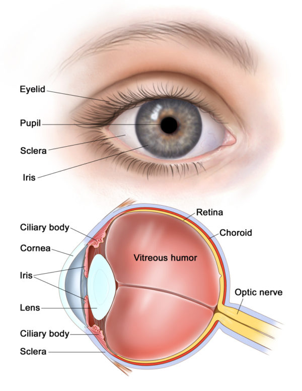
The human eye has components of the eyelid, eyelash, conjunctiva, sclera, cornea, pupil, iris, vitreous, lens (eye), auroral, retina and choroid.
Cornea
The cornea is a translucent, glass-like portion of the front of the eye, which is a cross section of an elliptical or an elliptical object, and beyond that the colored part of the eye, the iris, is seen. The cornea is composed of three main layers, which are the outer to the deep, respectively, the epithelium, stroma, and endothelium.
LASIK or laser surgeries are performed on this part to correct the number of glasses. With these surgeries, the corneal curvature is changed and the eye number is corrected.
Corneal transplantation is performed on diseases such as infections and dystrophies that cause corneal opacity and keratoconus that cause irregular corneal surface. In this type of surgery, the patient’s cornea will be replaced by a healthy human cornea that has died. Corneal transplantation is the most common and successful organ transplant in the human body.
Pupil
The pupil is actually a round hole in the center of the iris. The pupil resembles a photographic aperture and controls the amount of light entering the eye by dilating and shrinking the muscles within it.
Iris
The iris is the colored part of the eye that lies behind the cornea and is available in various colors such as blue, green, brown, honey and so on. Due to the transparency of the cornea, the iris can be seen well beyond it. The features and structure of every person’s iris are as unique as their fingerprints.
Aqueous liquid
It is a clear fluid that flows in front of the eye and between the cornea and the lens. This fluid delivers nutrients to the anterior parts of the eye. This fluid is always produced and discharged into the eye and subsequently discharged from the eye. Excessive accumulation within the eye, for whatever reason, increases intraocular pressure and subsequently damages the optic nerve and glaucoma.
Lens
The lens is located behind the pupil and performs the function of focusing and focusing light on the retina. The power of the lens in healthy individuals is about 20 diopters (the lens power unit). At an early age, the building has the ability to change power, making it possible to see distant and near-by objects in both images. This is called a lenses act. This ability decreases from the age of 45-40, and therefore, the eye becomes old, meaning that separate glasses will be required for close viewing and study.
Cataracts, one of the most common eye diseases, occur due to the opacity of the lens.
Extracorporeal muscles
The eye movement is performed by six muscles called the extracorporeal muscles. These include the outer straight muscle, the inner straight muscle, the superior straight and inferior straight muscle, and the tendon and upper and lower muscles.
sclera
Outer lining is a white that forms the wall of the eye behind and around the cornea.
choroid
The vascular layer is between the sclera and the retina, which is responsible for blood supply and nourishment of the posterior parts of the eye.
vitreous
The vitreous is actually a transparent jelly-like material that fills the area behind the lens. This gel-like substance has a higher consistency at younger ages and changes with this gel increase with age, which decreases its consistency. Changes in the vitreous gel are also more frequent in people who are close-knit because their back area is larger than others. These changes can be seen in the form of moving hair-like or fly wings in their field of vision. Especially when looking at a bright, white background, these shapes get more attention.
Due to the natural adhesion of the vitreous to the retina, when a new flywheel or luminous spark is created, the eye should be carefully examined to ensure that the retina is not ruptured or punctured.
Retina
The retina is a sensitive eye curtain that perfectly carpets the bottom of the eye. It receives images that enter the eye and converts them to nerve waves and transmits them through the optic nerve to the brain.
The center of vision or macula is a region of the center of the retina that is about 2.5 mm in diameter, with the highest degree of visual acuity concentrated in this part of the retina. If the area is damaged, vision can be reduced by up to 5%. Macular Degenerative Disease (FDA), which occurs in some people at an early age and causes moderate to severe vision loss, occurs in this area.
optic nerve
The optic nerve is in fact a retina of sensitive cells that reaches parts of the brain and transmits visual waves from the retina to the brain.
Diseases such as multiple sclerosis, or MS, can cause damage to the optic nerve and reduce vision. Toxins such as methyl alcohol (wood alcohol) can cause permanent damage to the optic nerve and dry out (atrophy) of the optic nerve and cause untreatable blindness.
Leave A Comment